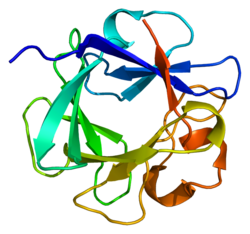
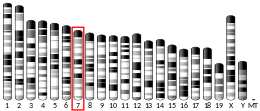
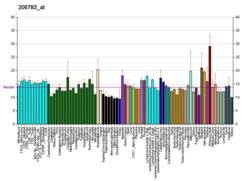
Fibroblast growth factor 4 is a protein that in humans is encoded by the FGF4 gene.[5][6]
The protein encoded by this gene is a member of the fibroblast growth factor (FGF) family. FGF family members possess broad mitogenic and cell survival activities and are involved in a variety of biological processes including embryonic development, cell growth, morphogenesis, tissue repair, tumor growth and invasion. This gene was identified by its oncogenic transforming activity. This gene and FGF3, another oncogenic growth factor, are located closely on chromosome 11. Co-amplification of both genes was found in various kinds of human tumors. Studies on the mouse homolog suggested a function in bone morphogenesis and limb development through the sonic hedgehog (SHH) signaling pathway.[6]
Function
[edit]During embryonic development, the 21-kD protein FGF4 functions as a signaling molecule that is involved in many important processes.[7][8] Studies using Fgf4 gene knockout mice showed developmental defects in embryos both in vivo and in vitro, revealing that FGF4 facilitates the survival and growth of the inner cell mass during the postimplantation phase of development by acting as an autocrine or paracrine ligand.[7] FGFs produced in the apical ectodermal ridge (AER) are critical for the proper forelimb and hindlimb outgrowth.[9] FGF signaling in the AER is involved in regulating limb digit number and cell death in the interdigital mesenchyme.[10] When FGF signaling dynamics and regulatory processes are altered, postaxial polydactyly and cutaneous syndactyly, two phenotypic abnormalities collectively known as polysyndactyly, can occur in the limbs. Polysyndactyly is observed when an excess of Fgf4 is expressed in limb buds of wild-type mice. In mutant limb buds that do not express Fgf8, the expression of Fgf4 still results in polysyndactyly, but Fgf4 is also able to rescue all skeletal defects that arise from the lack of Fgf8. Therefore, the Fgf4 gene compensates for the loss of the Fgf8 gene, revealing that FGF4 and FGF8 perform similar functions in limb skeleton patterning and limb development.[10] Studies of zebrafish Fgf4 knockdown embryos demonstrated that when Fgf4 signaling is inhibited, randomized left-right patterning of the liver, pancreas, and heart takes place, showing that Fgf4 is a crucial gene involved in developing left-right patterning of visceral organs. Furthermore, unlike the role of FGF4 in limb development, FGF4 and FGF8 have distinct roles and function independently in the process of visceral organ left-right patterning.[11] Fgf signaling pathway has also been demonstrated to drive hindgut identity during gastrointestinal development, and the up regulation of the Fgf4 in pluripotent stem cell has been used to direct their differentiation for the generation of intestinal Organoids and tissues in vitro.[12]
FGF4 Retrogenes
[edit]In canines the FGF4 retrogene insertion on chromosome 18 is involved in the short leg phenotype.[13] This is still a member of the FGF4 gene family. Fibroblast Growth Factor 4 is a protein coding gene, meaning it's a structural protein molecule.[14] The biological role that FGF4-18 plays is important in embryological development, specifically appropriate growth. In canines, the developmental structure this retrogene mutation patterning leads to is shortened legs due to the defects in endochondral ossification.These mutations and FGF signaling abnormalities are also linked in humans with dwarfism by preventing bones from growing to the normal length.[13][15] This FGF4 retrogene on not only chromosome 18 but also 12 leads to shortened limbs and abnormal vertebrae associated with intervertebral disc disease. Research done at University of California-Davis has found that FGF4 retrogene on chromosome 12 is also attributed to the short legs and abnormal intervertebral disc that degenerate.[13] This particular FGF4-12 retrogene in canines leads to the short limb phenotype from dysplastic shortened long bones, premature degeneration, and calcification of the intervertebral disc; which gives a susceptibility to IVDD (intervertebral disc disease).[13][15]
References
[edit]- ^ a b c GRCh38: Ensembl release 89: ENSG00000075388 – Ensembl, May 2017
- ^ a b c GRCm38: Ensembl release 89: ENSMUSG00000050917 – Ensembl, May 2017
- ^ "Human PubMed Reference:". National Center for Biotechnology Information, U.S. National Library of Medicine.
- ^ "Mouse PubMed Reference:". National Center for Biotechnology Information, U.S. National Library of Medicine.
- ^ Galland F, Stefanova M, Lafage M, Birnbaum D (Jul 1992). "Localization of the 5' end of the MCF2 oncogene to human chromosome 15q15----q23". Cytogenetics and Cell Genetics. 60 (2): 114–6. doi:10.1159/000133316. PMID 1611909.
- ^ a b "Entrez Gene: FGF4 fibroblast growth factor 4 (heparin secretory transforming protein 1, Kaposi sarcoma oncogene)".
- ^ a b Feldman B, Poueymirou W, Papaioannou VE, DeChiara TM, Goldfarb M (Jan 1995). "Requirement of FGF-4 for postimplantation mouse development". Science. 267 (5195): 246–9. Bibcode:1995Sci...267..246F. doi:10.1126/science.7809630. PMID 7809630. S2CID 31312392.
- ^ Yuan H, Corbi N, Basilico C, Dailey L (Nov 1995). "Developmental-specific activity of the FGF-4 enhancer requires the synergistic action of Sox2 and Oct-3". Genes & Development. 9 (21): 2635–45. doi:10.1101/gad.9.21.2635. PMID 7590241.
- ^ Boulet AM, Moon AM, Arenkiel BR, Capecchi MR (Sep 2004). "The roles of Fgf4 and Fgf8 in limb bud initiation and outgrowth". Developmental Biology. 273 (2): 361–72. doi:10.1016/j.ydbio.2004.06.012. PMID 15328019.
- ^ a b Lu P, Minowada G, Martin GR (Jan 2006). "Increasing Fgf4 expression in the mouse limb bud causes polysyndactyly and rescues the skeletal defects that result from loss of Fgf8 function". Development. 133 (1): 33–42. doi:10.1242/dev.02172. PMID 16308330.
- ^ Yamauchi H, Miyakawa N, Miyake A, Itoh N (Aug 2009). "Fgf4 is required for left-right patterning of visceral organs in zebrafish". Developmental Biology. 332 (1): 177–85. doi:10.1016/j.ydbio.2009.05.568. PMID 19481538.
- ^ Lancaster MA, Knoblich JA (2014). "Organogenesis in a dish: modeling development and disease using organoid technologies". Science. 345 (6194): 1247125. doi:10.1126/science.1247125. PMID 25035496. S2CID 16105729.
- ^ a b c d "Genetic Discovery Finds Dachshunds' Short-Leg Phenotype Linked To IVDD". www.purinaproclub.com. Retrieved 2022-04-13.
- ^ "FGFR3 Gene - GeneCards | FGFR3 Protein | FGFR3 Antibody". www.genecards.org. Retrieved 2022-04-13.
- ^ a b Brown EA, Dickinson PJ, Mansour T, Sturges BK, Aguilar M, Young AE, Korff C, Lind J, Ettinger CL, Varon S, Pollard R (2017-10-24). "FGF4 retrogene on CFA12 is responsible for chondrodystrophy and intervertebral disc disease in dogs". Proceedings of the National Academy of Sciences. 114 (43): 11476–11481. Bibcode:2017PNAS..11411476B. doi:10.1073/pnas.1709082114. ISSN 0027-8424. PMC 5664524. PMID 29073074.
Further reading
[edit]- Powers CJ, McLeskey SW, Wellstein A (Sep 2000). "Fibroblast growth factors, their receptors and signaling". Endocrine-Related Cancer. 7 (3): 165–97. CiteSeerX 10.1.1.323.4337. doi:10.1677/erc.0.0070165. PMID 11021964.
- Taira M, Yoshida T, Miyagawa K, Sakamoto H, Terada M, Sugimura T (May 1987). "cDNA sequence of human transforming gene hst and identification of the coding sequence required for transforming activity". Proceedings of the National Academy of Sciences of the United States of America. 84 (9): 2980–4. Bibcode:1987PNAS...84.2980T. doi:10.1073/pnas.84.9.2980. PMC 304784. PMID 2953031.
- Delli Bovi P, Curatola AM, Kern FG, Greco A, Ittmann M, Basilico C (Aug 1987). "An oncogene isolated by transfection of Kaposi's sarcoma DNA encodes a growth factor that is a member of the FGF family". Cell. 50 (5): 729–37. doi:10.1016/0092-8674(87)90331-X. PMID 2957062. S2CID 30018932.
- Yoshida T, Miyagawa K, Odagiri H, Sakamoto H, Little PF, Terada M, Sugimura T (Oct 1987). "Genomic sequence of hst, a transforming gene encoding a protein homologous to fibroblast growth factors and the int-2-encoded protein". Proceedings of the National Academy of Sciences of the United States of America. 84 (20): 7305–9. Bibcode:1987PNAS...84.7305Y. doi:10.1073/pnas.84.20.7305. PMC 299281. PMID 2959959.
- Wada A, Sakamoto H, Katoh O, Yoshida T, Yokota J, Little PF, Sugimura T, Terada M (Dec 1988). "Two homologous oncogenes, HST1 and INT2, are closely located in human genome". Biochemical and Biophysical Research Communications. 157 (2): 828–35. doi:10.1016/S0006-291X(88)80324-3. PMID 2974287.
- Huebner K, Ferrari AC, Delli Bovi P, Croce CM, Basilico C (1989). "The FGF-related oncogene, K-FGF, maps to human chromosome region 11q13, possibly near int-2". Oncogene Research. 3 (3): 263–70. PMID 3060803.
- Adelaide J, Mattei MG, Marics I, Raybaud F, Planche J, De Lapeyriere O, Birnbaum D (Apr 1988). "Chromosomal localization of the hst oncogene and its co-amplification with the int.2 oncogene in a human melanoma". Oncogene. 2 (4): 413–6. PMID 3283658.
- Ornitz DM, Xu J, Colvin JS, McEwen DG, MacArthur CA, Coulier F, Gao G, Goldfarb M (Jun 1996). "Receptor specificity of the fibroblast growth factor family". The Journal of Biological Chemistry. 271 (25): 15292–7. doi:10.1074/jbc.271.25.15292. PMID 8663044.
- Tanaka S, et al. (1998). "Promotion of trophoblast stem cell proliferation by FGF-4". Science. 282 (5396): 2072–2075. Bibcode:1998Sci...282.2072T. doi:10.1126/science.282.5396.2072. PMID 9851926.
- Helland R, Berglund GI, Otlewski J, Apostoluk W, Andersen OA, Willassen NP, Smalås AO (Jan 1999). "High-resolution structures of three new trypsin-squash-inhibitor complexes: a detailed comparison with other trypsins and their complexes". Acta Crystallographica Section D. 55 (Pt 1): 139–48. Bibcode:1999AcCrD..55..139H. doi:10.1107/S090744499801052X. PMID 10089404.
- Bellosta P, Iwahori A, Plotnikov AN, Eliseenkova AV, Basilico C, Mohammadi M (Sep 2001). "Identification of receptor and heparin binding sites in fibroblast growth factor 4 by structure-based mutagenesis". Molecular and Cellular Biology. 21 (17): 5946–57. doi:10.1128/MCB.21.17.5946-5957.2001. PMC 87313. PMID 11486033.
- Britto JA, Evans RD, Hayward RD, Jones BM (Dec 2001). "From genotype to phenotype: the differential expression of FGF, FGFR, and TGFbeta genes characterizes human cranioskeletal development and reflects clinical presentation in FGFR syndromes". Plastic and Reconstructive Surgery. 108 (7): 2026–39, discussion 2040–6. doi:10.1097/00006534-200112000-00030. PMID 11743396.
- Yamamoto H, Ochiya T, Tamamushi S, Toriyama-Baba H, Takahama Y, Hirai K, Sasaki H, Sakamoto H, Saito I, Iwamoto T, Kakizoe T, Terada M (Jan 2002). "HST-1/FGF-4 gene activation induces spermatogenesis and prevents adriamycin-induced testicular toxicity". Oncogene. 21 (6): 899–908. doi:10.1038/sj.onc.1205135. PMID 11840335. S2CID 12296298.
- Sieuwerts AM, Martens JW, Dorssers LC, Klijn JG, Foekens JA (Apr 2002). "Differential effects of fibroblast growth factors on expression of genes of the plasminogen activator and insulin-like growth factor systems by human breast fibroblasts". Thrombosis and Haemostasis. 87 (4): 674–83. doi:10.1055/s-0037-1613065. PMID 12008951. S2CID 13862591.
- Koh KR, Ohta K, Nakamae H, Hino M, Yamane T, Takubo T, Tatsumi N (Oct 2002). "Differential effects of fibroblast growth factor-4, epidermal growth factor and transforming growth factor-beta1 on functional development of stromal layers in acute myeloid leukemia". Leukemia Research. 26 (10): 933–8. doi:10.1016/S0145-2126(02)00033-4. PMID 12163055.
- Lopez-Sanchez C, Climent V, Schoenwolf GC, Alvarez IS, Garcia-Martinez V (Aug 2002). "Induction of cardiogenesis by Hensen's node and fibroblast growth factors". Cell and Tissue Research. 309 (2): 237–49. doi:10.1007/s00441-002-0567-2. PMID 12172783. S2CID 11783465.
- Wang P, Branch DR, Bali M, Schultz GA, Goss PE, Jin T (Oct 2003). "The POU homeodomain protein OCT3 as a potential transcriptional activator for fibroblast growth factor-4 (FGF-4) in human breast cancer cells". The Biochemical Journal. 375 (Pt 1): 199–205. doi:10.1042/BJ20030579. PMC 1223663. PMID 12841847.
- Reményi A, Lins K, Nissen LJ, Reinbold R, Schöler HR, Wilmanns M (Aug 2003). "Crystal structure of a POU/HMG/DNA ternary complex suggests differential assembly of Oct4 and Sox2 on two enhancers". Genes & Development. 17 (16): 2048–59. doi:10.1101/gad.269303. PMC 196258. PMID 12923055.
- Hirai K, Sasaki H, Yamamoto H, Sakamoto H, Kubota Y, Kakizoe T, Terada M, Ochiya T (Mar 2004). "HST-1/FGF-4 protects male germ cells from apoptosis under heat-stress condition". Experimental Cell Research. 294 (1): 77–85. doi:10.1016/j.yexcr.2003.11.012. PMID 14980503.
- Grigor'eva EV, Shevchenko AI, Mazurok NA, Elisaphenko EA, Zhelezova AI, Shilov AG, Dyban PA, Dyban AP, Noniashvili EM, Slobodyanyuk SY, Nesterova TB, Brockdorff N, Zakian SM (2009). "FGF4 independent derivation of trophoblast stem cells from the common vole". PLOS ONE. 4 (9): e7161. Bibcode:2009PLoSO...4.7161G. doi:10.1371/journal.pone.0007161. PMC 2744875. PMID 19777059.
- Boulet AM, Capecchi MR (Nov 2012). "Signaling by FGF4 and FGF8 is required for axial elongation of the mouse embryo". Developmental Biology. 371 (2): 235–45. doi:10.1016/j.ydbio.2012.08.017. PMC 3481862. PMID 22954964.


