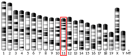
C-C chemokine receptor type 7 is a protein that in humans is encoded by the CCR7 gene.[5] Two ligands have been identified for this receptor: the chemokines (C-C motif) ligand 19 (CCL19/ELC) and (C-C motif) ligand 21 (CCL21).[6] The ligands have similar affinity for the receptor, though CCL19 has been shown to induce internalisation of CCR7 and desensitisation of the cell to CCL19/CCL21 signals.[7] CCR7 is a transmembrane protein with 7 transmembrane domains, which is coupled with heterotrimeric G proteins, which transduce the signal downstream through various signalling cascades. The main function of the receptor is to guide immune cells to immune organs (lymph nodes, thymus, spleen) by detecting specific chemokines, which these tissues secrete.[7]
CCR7 has also recently been designated CD197 (cluster of differentiation 197).
Function
[edit]The protein encoded by this gene is a member of the G protein-coupled receptor family. This receptor was identified as a gene induced by the Epstein–Barr virus (EBV), and is thought to be a mediator of EBV effects on B lymphocytes.[8] As stated above, the receptor guides immune cells to immune organs such as lymph nodes, which is needed for the development of both resistance and tolerance, but it is also important for development of T cells in thymus. The receptor is expressed mostly on adaptive immune cell types, namely thymocytes, naive T and B cells, regulatory T cells, central memory lymphocytes, but also dendritic cells.[7] CCR7 has been shown to stimulate dendritic cell maturation. CCR7 is also involved in homing of T cells to various secondary lymphoid organs such as lymph nodes and the spleen as well as trafficking of T cells within the spleen.[8]
CCR7 in dendritic cells
[edit]CCR7´s function is best studied in dendritic cells. Their activation in peripheral tissues induces CCR7 expression on the cell's surface, which recognize CCL19 and CCL21 produced in the Lymph node and increases dendritic cell expression of co-stimulation molecules (B7), and MHC class I or MHC class II.[9] CCR7 signalling was also found to affect chemotaxis, actin dynamics but also survival of dendritic cells, though all of the mentioned functions are induced by different independent signalling pathways.[10] Chemotaxis is regulated by MAPK pathway and surprisingly is independent of CCR7 signalling pathway regulating actin dynamics. Executive components of this cascade are kinases MEK1/2, ERK1/2, p38, JNK and perhaps others. The executive kinases phosphorylate transcription factors and other regulators thereby changing expression profile of the cell.[10] Increased cellular survival upon CCR7 ligation stems from both pro-apoptotic molecules inhibition and survival promoting proteins stimulation as the receptor is known to activate the PI3K/AKT/mTOR pathway The effector molecules of this pathway are mTOR and NFkB, collectively the effect is exerted via anti-apoptotic Bcl2 proteins expression and inhibition of pro-apoptotic proteins GSK3B, FOXO1/3 and 4EBP1. CCR7 affects cellular actin dynamics via the RhoA/cofilin pathway.[10]
Influence of CCR7 on central tolerance
[edit]CCR7 has been shown to be important for the selection process of T cells in thymus and its morphology formation. Experiments in mouse models have shown that mice lacking CCR7 had fewer thymocytes during development and more frequent autoimmune disorders. It is believed, that CCR7 takes part in homing of lymphoid progenitors to thymus, but also in thymocyte transition from thymic cortex to medulla.[7] Once double negative thymocyte (first step of T cell development) undergoes positive selection, it becomes double positive (expressing both CD4 and CD8 coreceptors) and starts to express CCR7, which guides it to thymic medulla, where negative selection takes place. ccr7 knockout mice have leaky negative selection are prone autoimmune disorders. The mechanism is thought to be both thymus morphology disruption and insufficient T cell receptor stimulation [7] It must however be noted that CCR7 affects not only central tolerance, but also peripheral tolerance by allowing homing of tolerogenic dendritic cells to lymph nodes.[11]
Clinical significance
[edit]CCR7 is expressed by various cancer cells, such as nonsmall lung cancer, gastric cancer and oesophageal cancer.[12][13][14] Expression of CCR7, usually with VEGF family proteins, by cancer cells is linked with metastasis and generally poorer prognosis.[15] Multiple mechanisms through which CCR7 expression changes the prognosis of cancer patients have been discovered.[16] As described above on the example of dendritic cells, CCR7 enhances survival of the cell and enables it to migrate following CCL19/CCL21 gradient, which leads to lymph nodes, in addition to that it has been shown that CCR7 ligation promotes EMT transition, which is cruicial for metastasis, as it allows cells to detach and migrate. Also CCR7 signalling induces VEGF-C and VEGF-D molecules, which promote lymphoneogenesis around the tumour.[16]
References
[edit]- ^ a b c GRCh38: Ensembl release 89: ENSG00000126353 – Ensembl, May 2017
- ^ a b c GRCm38: Ensembl release 89: ENSMUSG00000037944 – Ensembl, May 2017
- ^ "Human PubMed Reference:". National Center for Biotechnology Information, U.S. National Library of Medicine.
- ^ "Mouse PubMed Reference:". National Center for Biotechnology Information, U.S. National Library of Medicine.
- ^ Birkenbach M, Josefsen K, Yalamanchili R, Lenoir G, Kieff E (April 1993). "Epstein-Barr virus-induced genes: first lymphocyte-specific G protein-coupled peptide receptors". Journal of Virology. 67 (4): 2209–2220. doi:10.1128/JVI.67.4.2209-2220.1993. PMC 240341. PMID 8383238.
- ^ F. Balkwill, Cancer and the Chemokine Network, Nature reviews, 2004
- ^ a b c d e Alrumaihi F (2022). "The Multi-Functional Roles of CCR7 in Human Immunology and as a Promising Therapeutic Target for Cancer Therapeutics". Frontiers in Molecular Biosciences. 9: 834149. doi:10.3389/fmolb.2022.834149. PMC 9298655. PMID 35874608.
- ^ a b Sharma N, Benechet AP, Lefrançois L, Khanna KM (December 2015). "CD8 T Cells Enter the Splenic T Cell Zones Independently of CCR7, but the Subsequent Expansion and Trafficking Patterns of Effector T Cells after Infection Are Dysregulated in the Absence of CCR7 Migratory Cues". Journal of Immunology. 195 (11): 5227–5236. doi:10.4049/jimmunol.1500993. PMC 4655190. PMID 26500349.
- ^ Riol-Blanco L, Sánchez-Sánchez N, Torres A, Tejedor A, Narumiya S, Corbí AL, et al. (April 2005). "The chemokine receptor CCR7 activates in dendritic cells two signaling modules that independently regulate chemotaxis and migratory speed". Journal of Immunology. 174 (7): 4070–4080. doi:10.4049/jimmunol.174.7.4070. PMID 15778365.
- ^ a b c Rodríguez-Fernández JL, Criado-García O (2020). "The Chemokine Receptor CCR7 Uses Distinct Signaling Modules With Biased Functionality to Regulate Dendritic Cells". Frontiers in Immunology. 11: 528. doi:10.3389/fimmu.2020.00528. PMC 7174648. PMID 32351499.
- ^ Brandum EP, Jørgensen AS, Rosenkilde MM, Hjortø GM (August 2021). "Dendritic Cells and CCR7 Expression: An Important Factor for Autoimmune Diseases, Chronic Inflammation, and Cancer". International Journal of Molecular Sciences. 22 (15): 8340. doi:10.3390/ijms22158340. PMC 8348795. PMID 34361107.
- ^ Mashino K, Sadanaga N, Yamaguchi H, Tanaka F, Ohta M, Shibuta K, et al. (May 2002). "Expression of chemokine receptor CCR7 is associated with lymph node metastasis of gastric carcinoma". Cancer Research. 62 (10): 2937–2941. PMID 12019175.
- ^ Takanami I (June 2003). "Overexpression of CCR7 mRNA in nonsmall cell lung cancer: correlation with lymph node metastasis". International Journal of Cancer. 105 (2): 186–189. doi:10.1002/ijc.11063. PMID 12673677. S2CID 1523901.
- ^ Ding Y, Shimada Y, Maeda M, Kawabe A, Kaganoi J, Komoto I, et al. (August 2003). "Association of CC chemokine receptor 7 with lymph node metastasis of esophageal squamous cell carcinoma". Clinical Cancer Research. 9 (9): 3406–3412. PMID 12960129.
- ^ Shields JD, Fleury ME, Yong C, Tomei AA, Randolph GJ, Swartz MA (June 2007). "Autologous chemotaxis as a mechanism of tumor cell homing to lymphatics via interstitial flow and autocrine CCR7 signaling". Cancer Cell. 11 (6): 526–538. doi:10.1016/j.ccr.2007.04.020. PMID 17560334.
- ^ a b Salem A, Alotaibi M, Mroueh R, Basheer HA, Afarinkia K (January 2021). "CCR7 as a therapeutic target in Cancer". Biochimica et Biophysica Acta (BBA) - Reviews on Cancer. 1875 (1): 188499. doi:10.1016/j.bbcan.2020.188499. PMID 33385485. S2CID 230108218.
External links
[edit]- Human CCR7 genome location and CCR7 gene details page in the UCSC Genome Browser.
Further reading
[edit]- Müller G, Lipp M (June 2003). "Shaping up adaptive immunity: the impact of CCR7 and CXCR5 on lymphocyte trafficking". Microcirculation. 10 (3–4): 325–334. doi:10.1038/sj.mn.7800197. PMID 12851649. S2CID 1785143.
- Burgstahler R, Kempkes B, Steube K, Lipp M (October 1995). "Expression of the chemokine receptor BLR2/EBI1 is specifically transactivated by Epstein-Barr virus nuclear antigen 2". Biochemical and Biophysical Research Communications. 215 (2): 737–743. doi:10.1006/bbrc.1995.2525. PMID 7488016.
- Schweickart VL, Raport CJ, Godiska R, Byers MG, Eddy RL, Shows TB, Gray PW (October 1994). "Cloning of human and mouse EBI1, a lymphoid-specific G-protein-coupled receptor encoded on human chromosome 17q12-q21.2". Genomics. 23 (3): 643–650. doi:10.1006/geno.1994.1553. PMID 7851893.
- Bonaldo MF, Lennon G, Soares MB (September 1996). "Normalization and subtraction: two approaches to facilitate gene discovery". Genome Research. 6 (9): 791–806. doi:10.1101/gr.6.9.791. PMID 8889548.
- Yoshida R, Imai T, Hieshima K, Kusuda J, Baba M, Kitaura M, et al. (May 1997). "Molecular cloning of a novel human CC chemokine EBI1-ligand chemokine that is a specific functional ligand for EBI1, CCR7". The Journal of Biological Chemistry. 272 (21): 13803–13809. doi:10.1074/jbc.272.21.13803. PMID 9153236.
- Yoshida R, Nagira M, Kitaura M, Imagawa N, Imai T, Yoshie O (March 1998). "Secondary lymphoid-tissue chemokine is a functional ligand for the CC chemokine receptor CCR7". The Journal of Biological Chemistry. 273 (12): 7118–7122. doi:10.1074/jbc.273.12.7118. PMID 9507024.
- Campbell JJ, Bowman EP, Murphy K, Youngman KR, Siani MA, Thompson DA, et al. (May 1998). "6-C-kine (SLC), a lymphocyte adhesion-triggering chemokine expressed by high endothelium, is an agonist for the MIP-3beta receptor CCR7". The Journal of Cell Biology. 141 (4): 1053–1059. doi:10.1083/jcb.141.4.1053. PMC 2132769. PMID 9585422.
- Kim CH, Pelus LM, White JR, Broxmeyer HE (September 1998). "Macrophage-inflammatory protein-3 beta/EBI1-ligand chemokine/CK beta-11, a CC chemokine, is a chemoattractant with a specificity for macrophage progenitors among myeloid progenitor cells". Journal of Immunology. 161 (5): 2580–2585. doi:10.4049/jimmunol.161.5.2580. PMID 9725259. S2CID 255361893.
- Yanagihara S, Komura E, Nagafune J, Watarai H, Yamaguchi Y (September 1998). "EBI1/CCR7 is a new member of dendritic cell chemokine receptor that is up-regulated upon maturation". Journal of Immunology. 161 (6): 3096–3102. doi:10.4049/jimmunol.161.6.3096. PMID 9743376. S2CID 26084149.
- Hasegawa H, Nomura T, Kohno M, Tateishi N, Suzuki Y, Maeda N, et al. (January 2000). "Increased chemokine receptor CCR7/EBI1 expression enhances the infiltration of lymphoid organs by adult T-cell leukemia cells". Blood. 95 (1): 30–38. doi:10.1182/blood.V95.1.30. PMID 10607681.
- Gosling J, Dairaghi DJ, Wang Y, Hanley M, Talbot D, Miao Z, Schall TJ (March 2000). "Cutting edge: identification of a novel chemokine receptor that binds dendritic cell- and T cell-active chemokines including ELC, SLC, and TECK". Journal of Immunology. 164 (6): 2851–2856. doi:10.4049/jimmunol.164.6.2851. PMID 10706668.
- Agace WW, Roberts AI, Wu L, Greineder C, Ebert EC, Parker CM (March 2000). "Human intestinal lamina propria and intraepithelial lymphocytes express receptors specific for chemokines induced by inflammation". European Journal of Immunology. 30 (3): 819–826. doi:10.1002/1521-4141(200003)30:3<819::AID-IMMU819>3.0.CO;2-Y. PMID 10741397.
- Annunziato F, Romagnani P, Cosmi L, Beltrame C, Steiner BH, Lazzeri E, et al. (July 2000). "Macrophage-derived chemokine and EBI1-ligand chemokine attract human thymocytes in different stage of development and are produced by distinct subsets of medullary epithelial cells: possible implications for negative selection". Journal of Immunology. 165 (1): 238–246. doi:10.4049/jimmunol.165.1.238. PMID 10861057.
- Campbell JJ, Murphy KE, Kunkel EJ, Brightling CE, Soler D, Shen Z, et al. (January 2001). "CCR7 expression and memory T cell diversity in humans". Journal of Immunology. 166 (2): 877–884. doi:10.4049/jimmunol.166.2.877. PMID 11145663.
- Campbell JJ, Qin S, Unutmaz D, Soler D, Murphy KE, Hodge MR, et al. (June 2001). "Unique subpopulations of CD56+ NK and NK-T peripheral blood lymphocytes identified by chemokine receptor expression repertoire". Journal of Immunology. 166 (11): 6477–6482. doi:10.4049/jimmunol.166.11.6477. PMID 11359797.
- Taylor JR, Kimbrell KC, Scoggins R, Delaney M, Wu L, Camerini D (September 2001). "Expression and function of chemokine receptors on human thymocytes: implications for infection by human immunodeficiency virus type 1". Journal of Virology. 75 (18): 8752–8760. doi:10.1128/JVI.75.18.8752-8760.2001. PMC 115120. PMID 11507220.
- Höpken UE, Foss HD, Meyer D, Hinz M, Leder K, Stein H, Lipp M (February 2002). "Up-regulation of the chemokine receptor CCR7 in classical but not in lymphocyte-predominant Hodgkin disease correlates with distinct dissemination of neoplastic cells in lymphoid organs". Blood. 99 (4): 1109–1116. doi:10.1182/blood.V99.4.1109. PMID 11830455.
- Nakayama T, Fujisawa R, Izawa D, Hieshima K, Takada K, Yoshie O (March 2002). "Human B cells immortalized with Epstein-Barr virus upregulate CCR6 and CCR10 and downregulate CXCR4 and CXCR5". Journal of Virology. 76 (6): 3072–3077. doi:10.1128/JVI.76.6.3072-3077.2002. PMC 135988. PMID 11861876.
- Till KJ, Lin K, Zuzel M, Cawley JC (April 2002). "The chemokine receptor CCR7 and alpha4 integrin are important for migration of chronic lymphocytic leukemia cells into lymph nodes". Blood. 99 (8): 2977–2984. doi:10.1182/blood.V99.8.2977. PMID 11929789.
- Sánchez-Sánchez N, Riol-Blanco L, de la Rosa G, Puig-Kröger A, García-Bordas J, Martín D, et al. (August 2004). "Chemokine receptor CCR7 induces intracellular signaling that inhibits apoptosis of mature dendritic cells". Blood. 104 (3): 619–625. doi:10.1182/blood-2003-11-3943. PMID 15059845.
- López-Cotarelo P, Escribano-Díaz C, González-Bethencourt IL, Gómez-Moreira C, Deguiz ML, Torres-Bacete J, et al. (January 2015). "A novel MEK-ERK-AMPK signaling axis controls chemokine receptor CCR7-dependent survival in human mature dendritic cells". The Journal of Biological Chemistry. 290 (2): 827–840. doi:10.1074/jbc.M114.596551. PMC 4294505. PMID 25425646.
- Riol-Blanco L, Sánchez-Sánchez N, Torres A, Tejedor A, Narumiya S, Corbí AL, et al. (April 2005). "The chemokine receptor CCR7 activates in dendritic cells two signaling modules that independently regulate chemotaxis and migratory speed". Journal of Immunology. 174 (7): 4070–4080. doi:10.4049/jimmunol.174.7.4070. PMID 15778365.
- Torres-Bacete J, Delgado-Martín C, Gómez-Moreira C, Simizu S, Rodríguez-Fernández JL (August 2015). "The Mammalian Sterile 20-like 1 Kinase Controls Selective CCR7-Dependent Functions in Human Dendritic Cells". Journal of Immunology. 195 (3): 973–981. doi:10.4049/jimmunol.1401966. PMID 26116501.
- Sánchez-Sánchez N, Riol-Blanco L, Rodríguez-Fernández JL (May 2006). "The multiple personalities of the chemokine receptor CCR7 in dendritic cells". Journal of Immunology. 176 (9): 5153–5159. doi:10.4049/jimmunol.176.9.5153. PMID 16621978.
- Escribano C, Delgado-Martín C, Rodríguez-Fernández JL (November 2009). "CCR7-dependent stimulation of survival in dendritic cells involves inhibition of GSK3beta". Journal of Immunology. 183 (10): 6282–6295. doi:10.4049/jimmunol.0804093. PMID 19841191.
This article incorporates text from the United States National Library of Medicine, which is in the public domain.


